

Patients with colonic IBD are at increased risk of colorectal cancer, with an additional challenge: dysplasia is often subtle, flat, and hidden among post-inflammatory mucosal changes. Historically, the answer has been dye-based chromoendoscopy, recommended to improve dysplasia detection. But in the era of high-definition imaging and virtual chromoendoscopy, is the dye itself really what makes the difference?
A meta-analysis by Mohamed et al., including 6 randomized trials and 978 patients, confirmed an advantage of dye-based chromoendoscopy over HD white-light endoscopy for dysplasia detection (OR 1.95; 95% CI 1.21–3.11).
However, HELIOS challenges this interpretation. In this randomized trial published in Gut, HD white-light endoscopy with segmental re-inspection was non-inferior to standard dye-based chromoendoscopy for neoplasia detection (10.3% vs 13.1%). Importantly, 19% of all lesions were detected only during the second inspection. Withdrawal time remained shorter with white-light endoscopy (19 vs 26 min), but neoplasia yield per 10 minutes of inspection was virtually identical (0.0616 vs 0.0584; p=0.83).
So, is it about better contrast—or simply better inspection?
Virtual chromoendoscopy may provide an attractive compromise. A recent meta-analysis of 8 studies found no significant difference in neoplasia detection compared with dye-based chromoendoscopy (OR 1.20; 95% CI 0.86–1.70).
But perhaps the key message lies elsewhere.
You only find what you are looking for — and you need to give yourself enough time to find it.
In Barrett’s esophagus, guidelines have already introduced the concept of a minimum inspection time per centimeter of Barrett’s mucosa. Perhaps IBD surveillance should similarly consider inspection time and inspection quality as key quality indicators.
Optimal bowel preparation, a slowly and carefully inspected colon, ideally during disease remission, and above all an endoscopist trained to recognize the sometimes extremely subtle features of IBD-associated dysplasia may ultimately matter more than the choice between methylene blue, indigo carmine, NBI, BLI, or white-light imaging.
The best chromoendoscopy in the world will never compensate for an examination performed too quickly.
Improving the detection of dysplastic lesions during IBD surveillance does not currently require another new technology, but something much simpler: better bowel preparation, better inspection… and, above all, taking the time to look.
FROM THE ENDOGASTROLIVE MEDIA LIBRARY
Endoscopy in IBD: a closer look
(Please note that the following videos are currently available in French only)
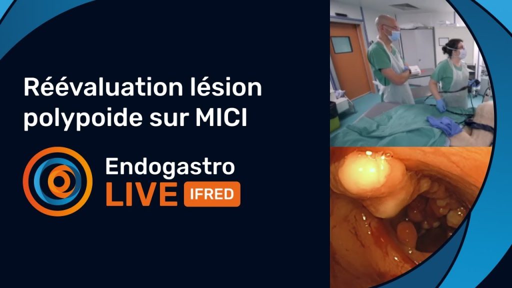
Reassessment of a Polypoid Lesion in IBD
Watch the video ›
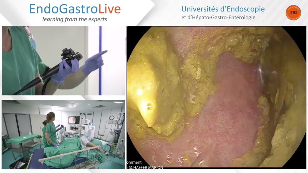
Endoscopic Assessment of Ulcerative Colitis (UC)
Watch the video ›
References
- Dye Chromoendoscopy Outperforms High-Definition White Light Endoscopy in Dysplasia Detection for Patients With Inflammatory Bowel Disease: An Updated Meta-Analysis of Randomized Controlled Trials
Mohamed MFH, Marino D, Elfert K, Beran A, Nayfeh T, Abdallah MA, et al. Am J Gastroenterol. 2024 Apr 1;119(4):719-726. doi: 10.14309/ajg.0000000000002595. - Surveillance in inflammatory bowel disease: white light endoscopy with segmental re-inspection versus dye-based chromoendoscopy – a multi-arm randomised controlled trial (HELIOS)
Te Groen M, Wijnands AM, den Broeder N, de Jong DJ, van Dop WA, Duijvestein M, et al. Gut. 2025 Mar 6;74(4):547-556. doi: 10.1136/gutjnl-2024-333446. - Dysplasia detection using high-definition scopes with dye vs virtual chromoendoscopy in IBD: A meta-analysis of randomized clinical trials
Mohamed MFH, Chu JJ, Beran A, Nayfeh T, Ahmed K, Shah SA. Inflamm Bowel Dis. 2026 Jul 1;32(7):1410-1414. doi: 10.1093/ibd/izag019.
EndoGastroLive – The Scientific Newsletters
Endoscopy is both an art and a science. Beyond mastering techniques and procedures, practice relies on evidence that keeps evolving — and that deserves more than a quick read of an abstract.
The EndoGastroLive Scientific Newsletters offer short, punchy, critical analyses of the publications that matter in endoscopy and gastroenterology: not just the results, but their significance, their limitations, and whether — or not — they should change what we do tomorrow.
The science behind the scope.